
| Thoracic aorta | |
|---|---|
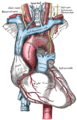
 Schematic view of the aorta, showing major branches and segments with thoracic aorta shown as continuation of descending aorta | |
 The thoracic aorta, viewed from the left side. | |
| Details | |
| Source | Descending aorta |
| Branches | Bronchial arteries, esophageal arteries, posterior intercostal arteries, abdominal aorta, superior phrenic artery, pericardial arteries |
| Identifiers | |
| Latin | aorta thoracalis |
| MeSH | D001013 |
| TA98 | A12.2.11.001 |
| TA2 | 4186 |
| FMA | 3786 |
| Anatomical terminology | |
The thoracic aorta is a part of the aorta located in the thorax. It is a continuation of the aortic arch. It is located within the posterior mediastinal cavity, but frequently bulges into the left pleural cavity. The descending thoracic aorta begins at the lower border of the fourth thoracic vertebra and ends in front of the lower border of the twelfth thoracic vertebra, at the aortic hiatus in the diaphragm where it becomes the abdominal aorta.
At its commencement, it is situated on the left of the vertebral column; it approaches the median line as it descends; and, at its termination, lies directly in front of the column.
The thoracic aorta has a curved shape that faces forward, and has small branches. It has a radius of approximately 1.16 cm.[1]
YouTube Encyclopedic
-
1/3Views:34 64838 7131 834
-
Thoracic (Descending) Aorta - Anatomy and its Branches - Human Anatomy | Kenhub
-
Traumatic Thoracic Aortic Aneurysm Repair Part 1 of 2
-
(46) Thorax د. أحمد كمال - Divisions of the thoracic aorta.data
Transcription
Hello everyone! This is Matt from Kenhub, and this tutorial will describe the thoracic aorta and its branches. The aorta seen here, highlighted in green, is the largest blood vessel in the body, and almost all arteries stem from this vessel or from one of its branches. Our focus in this video is a section of the aorta called the thoracic aorta or descending aorta and its branches. You can see the clear view of it here without any organs in the way. The terms thoracic aorta and descending aorta are used interchangeably, and they're both perfectly acceptable. Here's an overview of the aorta so that you can find your bearings when we discuss the specifics of the thoracic aorta. After starting at the aortic valve, the ascending aorta winds towards the head, becomes the aortic arch or the transverse aorta as it makes a rainbow over the superior aspect of the heart, and then moves in an inferior direction through the chest and abdomen. At the left subclavian artery, it becomes the thoracic aorta. After the thoracic aorta, comes the abdominal aorta and follows, then, the thoracoabdominal aorta which ends in its bifurcation into the left and right common iliac arteries. Let's now focus on the thoracic aorta. It begins at the level of the fourth thoracic vertebra and descends on the left side of the thoracic vertebrae from the fifth thoracic vertebra to the 12th thoracic vertebra. Running behind the base of the left lung and the pericardium, it enters the abdomen via the aortic hiatus of the diaphragm when it reaches the T12 vertebra. The branches of the thoracic aorta include the bronchial arteries, the pericardial arteries, the superior phrenic arteries, the esophageal arteries, the posterior intercostals arteries, and the subcostal arteries. Once again, those branches and the parts they supply: the bronchial which supplies the lungs, the pericardial which supplies the dorsal portion of the pericardium, the superior phrenic supporting the diaphragm and the adrenal glands, the esophageal supplying the (you guessed it!) the esophagus. The posterior intercostal, which you see here in green, supply the intercostal spaces and the subcostal, the flat abdominal wall muscles. This video is more fun than reading a textbook, right? If you want more videos, interactive quizzes, articles, and an atlas of human anatomy, click on the "Take me to Kenhub" button. It is time to say goodbye to your old textbooks and say hello to your new anatomy learning partner, Kenhub! See you there! https://www.kenhub.com
Structure
The thoracic aorta is part of the descending aorta, which has different parts named according to their structure or location. The thoracic aorta is a continuation of the descending aorta and becomes the abdominal aorta when it passes through the diaphragm. The initial part of the aorta, the ascending aorta, rises out of the left ventricle, from which it is separated by the aortic valve. The two coronary arteries of the heart arise from the aortic root, just above the cusps of the aortic valve. The aorta then arches back over the right pulmonary artery. Three vessels come out of the aortic arch: the brachiocephalic artery, the left common carotid artery, and the left subclavian artery. These vessels supply blood to the head, neck, thorax and upper limbs.
Behind the descending thoracic aorta is the vertebral column and the hemiazygos vein. To the right is the azygos veins and thoracic duct, and to the left is the left pleura and lung. In front of the thoracic aorta lies the root of the left lung, the pericardium, the esophagus, and the diaphragm.
The esophagus, which is covered by a nerve plexus lies to the right of the descending thoracic aorta. Lower, the esophagus passes in front of the aorta, and ultimately is situated on the left.
Function
The aorta is an artery that conveys oxygenated blood from the heart to other parts of the body. It is one of the largest arteries in the body.[2] The aorta gives off several paired branches as it descends. In descending order, these include the
- Bronchial arteries
- Mediastinal arteries
- Esophageal arteries
- Pericardial arteries
- Superior phrenic arteries
Note: The posterior intercostal arteries are branches that originate throughout the length of the posterior aspect of the descending thoracic aorta.
Clinical significance

Additional images
-
 Transverse section of thorax, showing relations of pulmonary artery.
 -
 The arch of the aorta, and its branches.


References
![]() This article incorporates text in the public domain from page 598 of the 20th edition of Gray's Anatomy (1918)
This article incorporates text in the public domain from page 598 of the 20th edition of Gray's Anatomy (1918)
- ^ Solutions to the Van der Pol Equation: a Model of Aortic Blood Flow
- ^ "Aorta Anatomy". UF Health, University of Florida Health. 2013-08-08. Retrieved 2022-06-05.
External links
| National | |
|---|---|
| Other | |