
| Tensor veli palatini muscle | |
|---|---|
 Dissection of the muscles of the palate from behind (tensor veli palatini visible at upper right). | |
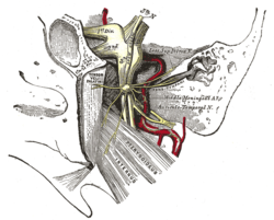 The otic ganglion and its branches (tensor veli palatini visible at center left). | |
| Details | |
| Origin | Medial pterygoid plate of the sphenoid bone (scaphoid fossa) |
| Insertion | Palatine aponeurosis |
| Nerve | Mandibular nerve (V3) |
| Actions | Tension of the soft palate |
| Identifiers | |
| Latin | musculus tensor veli palatini |
| TA98 | A05.2.01.103 |
| TA2 | 2129 |
| FMA | 46730 |
| Anatomical terms of muscle | |
The tensor veli palatini muscle (tensor palati or tensor muscle of the velum palatinum) is a thin, triangular muscle of the head that tenses the soft palate and opens the Eustachian tube to equalise pressure in the middle ear.
YouTube Encyclopedic
-
1/3Views:1 441167 9131 827
-
The veli palatini muscles
-
Soft palate: Muscles, Function & Definition - Human Anatomy | Kenhub
-
Muscles of soft palate-Tensor veli palatini
Transcription
Structure
The tensor veli palatini muscle is thin and triangular in shape.[1]
Origin
It arises from the scaphoid fossa of the pterygoid process of the sphenoid[1] anteriorly[citation needed], the (medial aspect of the) spine of sphenoid bone posteriorly, and - between the aforementioned anterior and posterior attachments - from the anterolateral aspect of the membranous wall of the pharyngotympanic tube.[1]
At the muscle's origin, some of its muscle fibres may be continuous with those of the tensor tympani muscle.[1]
Insertion
Inferiorly, the muscle converges to form a tendon of attachment. This tendon winds medially around the pterygoid hamulus (with a small bursa interposed between the two) to insert into the palatine aponeurosis and into the bony surface posterior to the palatine crest of the horizontal plate of palatine bone.[1]
Dilator tubae component
Some of the muscle's fibres insert onto the lateral lamina of the cartilaginous part of pharyngotympanic tube and adjacent connective tissue, and the Ostmann's fat pad.[1]
The portion of the muscle with these attachments is sometimes called the dilator tubae.[1]
Innervation
The tensor veli palatini muscle receives motor innervation from the mandibular nerve (CN V3) (a branch of the trigeminal nerve (CN V))[2] via the nerve to medial pterygoid.[1]
It is the only muscle of the palate not innervated by the pharyngeal plexus, which is formed by the vagal and glossopharyngeal nerves.[citation needed]
Relations
It is situated anterolaterally to the levator veli palatini muscle.[citation needed]
From its origin to its insertion, the muscle passes vertically between the medial pterygoid plate and the medial pterygoid muscle.[citation needed]
Actions/movements
Bilateral contraction of the two tensor veli palatini muscles makes the soft palate (especially its anterior portion) taut, as well as flattening the arch of the soft palate and thereby depressing it.[1]
Unilateral contraction draws the soft palate ipsilaterally.[1]
Function
The tensor veli palatini tenses the soft palate and by doing so, assists the levator veli palatini in elevating the palate to occlude and prevent entry of food into the nasopharynx during swallowing. The tensed palate consequently provides a stable platform for elevation of the pharynx during swallowing by the pharyngeal muscles.
Since it is also attached to the lateral cartilaginous lamina of the pharyngotympanic tube (auditory tube or Eustachian tube), it assists in its opening during swallowing or yawning to allow air pressure to equalize between the tympanic cavity and the outside air. Equalization of air pressure in the tympanic cavity is essential for preventing damage to the tympanic membrane and a resulting loss of hearing acuity.
Additional images
-
 Mandibular division of trigeminal nerve, seen from the middle line.

See also
References
![]() This article incorporates text in the public domain from page 1139 of the 20th edition of Gray's Anatomy (1918)
This article incorporates text in the public domain from page 1139 of the 20th edition of Gray's Anatomy (1918)
- ^ a b c d e f g h i j Standring, Susan (2020). Gray's Anatomy: The Anatomical Basis of Clinical Practice (42th ed.). New York. pp. 709–710. ISBN 978-0-7020-7707-4. OCLC 1201341621.
{{cite book}}: CS1 maint: location missing publisher (link) - ^ Fillmore, Erin P.; Seifert, Mark F. (2015). "22 - Anatomy of the Trigeminal Nerve". Nerves and Nerve Injuries. Vol. 1: History, Embryology, Anatomy, Imaging, and Diagnostics. Academic Press. pp. 319–350. doi:10.1016/B978-0-12-410390-0.00023-8. ISBN 978-0-12-410390-0.