
| Spinal canal | |
|---|---|
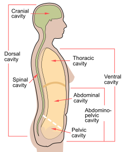 Spinal cavity shown as part of dorsal body cavity. | |
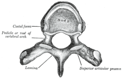 A typical thoracic vertebra viewed from above. (Spinal canal is not labeled, but the foramen in the center would make up part of it.) | |
| Details | |
| Identifiers | |
| Latin | c. vertebralis |
| MeSH | D013115 |
| TA98 | A02.2.00.009 |
| TA2 | 1009 |
| FMA | 9680 |
| Anatomical terminology | |
In human anatomy, the spinal canal, vertebral canal or spinal cavity is an elongated body cavity enclosed within the dorsal bony arches of the vertebral column, which contains the spinal cord, spinal roots and dorsal root ganglia. It is a process of the dorsal body cavity formed by alignment of the vertebral foramina. Under the vertebral arches, the spinal canal is also covered anteriorly by the posterior longitudinal ligament and posteriorly by the ligamentum flavum. The potential space between these ligaments and the dura mater covering the spinal cord is known as the epidural space. Spinal nerves exit the spinal canal via the intervertebral foramina under the corresponding vertebral pedicles.
In humans, the spinal cord gets outgrown by the vertebral column during development into adulthood, and the lower section of the spinal canal is occupied by the filum terminale and a bundle of spinal nerves known as the cauda equina instead of the actual spinal cord, which finishes at the L1/L2 level.
YouTube Encyclopedic
-
1/5Views:202 213141 11223 633393 47160 642
-
Lumbar Spinal Stenosis - What is the cause and how will I get better?
-
Symptoms of Cervical Stenosis | Jeffrey Cantor, MD
-
What Is Lumbar Lateral Stenosis? | Foraminal Stenosis
-
Bulging Disc
-
Spine Anatomy | Know Your Spine
Transcription
Structure
The vertebral canal is enclosed anteriorly by the vertebral bodies, intervertebral discs, and the posterior longitudinal ligament; it is enclosed posteriorly by the vertebral laminae and the ligamenta flava; laterally, it is incompletely enclosed by the pedicles with the interval between two adjacent pedicles on either side creating an intervertebral foramen (allowing the passage of the spinal nerves and radicular blood vessels).[1]
The vertebral canal progressively narrows inferiorly.[1] It is wider in the cervical region to accommodate the cervical enlargement of the spinal cord.[2][3]
Contents
The outermost layer of the meninges, the dura mater, is closely associated with the arachnoid mater which in turn is loosely connected to the innermost layer, the pia mater. The meninges divide the spinal canal into the epidural space and the subarachnoid space. The pia mater is closely attached to the spinal cord. A subdural space is generally only present due to trauma and/or pathological situations. The subarachnoid space is filled with cerebrospinal fluid and contains the vessels that supply the spinal cord, namely the anterior spinal artery and the paired posterior spinal arteries, accompanied by corresponding spinal veins. The anterior and posterior spinal arteries form anastomoses known as the vasocorona of the spinal cord and these supply nutrients to the canal. The epidural space contains loose fatty tissue, and a network of large, thin-walled blood vessels called the internal vertebral venous plexuses.[citation needed]
Clinical significance
Spinal stenosis is a narrowing of the canal which can occur in any region of the spine and can be caused by a number of factors. It may be caused by cervical myelopathy.[4]
Spinal canal endoscopy can be used to investigate the epidural space, and is an important spinal diagnostic technique.[5][6]
History
The spinal canal was first described by Jean Fernel.[citation needed]
References
- ^ a b Sinnatamby, Chummy S. (2011). Last's Anatomy (12th ed.). p. 425. ISBN 978-0-7295-3752-0.
- ^ Kim, Hak-Jin (2010-01-01), Kim, Daniel H.; Kim, Yong-Chul; Kim, Kyung-Hoon (eds.), "Chapter 3 - Radiologic Anatomy of the Spine", Minimally Invasive Percutaneous Spinal Techniques, New York: W.B. Saunders, pp. 46–57, doi:10.1016/b978-0-7020-2913-4.00003-3, ISBN 978-0-7020-2913-4, retrieved 2020-11-03
- ^ Haran, Crishan. "Spinal canal | Radiology Reference Article | Radiopaedia.org". Radiopaedia.
- ^ Lewit, Karel; Ellis, Richard M (2010-01-01), Lewit, Karel; Ellis, Richard M (eds.), "Chapter 3 - Functional anatomy and radiology of the spinal column", Manipulative Therapy, Edinburgh: Churchill Livingstone, pp. 39–85, doi:10.1016/b978-0-7020-3056-7.00003-6, ISBN 978-0-7020-3056-7, retrieved 2020-11-03
- ^ Datta, Sukdeb (2009-01-01), Smith, HOWARD S. (ed.), "Chapter 87 - EPIDURAL ADHESIOLYSIS", Current Therapy in Pain, Philadelphia: W.B. Saunders, pp. 629–639, doi:10.1016/b978-1-4160-4836-7.00087-0, ISBN 978-1-4160-4836-7, retrieved 2020-11-03
- ^ Saberski, Lloyd R. (2007-01-01), Waldman, Steven D.; Bloch, Joseph I. (eds.), "chapter 15 - Spinal Canal Endoscopy", Pain Management, Philadelphia: W.B. Saunders, pp. 167–178, doi:10.1016/b978-0-7216-0334-6.50019-4, ISBN 978-0-7216-0334-6, retrieved 2020-11-03
External links
| National | |
|---|---|
| Other | |