

| |
| |
| Names | |
|---|---|
| IUPAC name
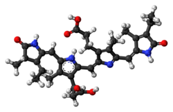
3-[(2Z,5E)-2-[[3-(2-carboxyethyl)-5-[(Z)-[(3R,4R)-3-ethyl-4-methyl-5-oxopyrrolidin-2-ylidene]methyl]-4-methyl-1H-pyrrol-2-yl]methylidene]-5-[(4-ethyl-3-methyl-5-oxopyrrol-2-yl)methylidene]-4-methylpyrrol-3-yl]propanoic acid
| |
| Identifiers | |
3D model (JSmol)
|
|
| 4285356 | |
| ChEBI | |
| ChemSpider | |
PubChem CID
|
|
| UNII | |
| |
| |
| Properties | |
| C33H38N4O6 | |
| Molar mass | 586.69 g/mol |
Except where otherwise noted, data are given for materials in their standard state (at 25 °C [77 °F], 100 kPa).
| |
Phycocyanobilin is a blue phycobilin, i.e., a tetrapyrrole chromophore found in cyanobacteria and in the chloroplasts of red algae, glaucophytes, and some cryptomonads. Phycocyanobilin is present only in the phycobiliproteins allophycocyanin and phycocyanin, of which it is the terminal acceptor of energy. It is covalently linked to these phycobiliproteins by a thioether bond.
Phycocyanobilin (PCB), has the ability to bind to human serum albumin (HSA), protein found mainly in the blood of humans. This PCB-HCA complex benefits the structure of HSA, increasing the thermal stability of HSA, as well as increasing its ability to prevent against proteolytic activity of other proteins.[1]
YouTube Encyclopedic
-
1/1Views:2 518
-
Structural Studies of Phycobiliproteins from Spirulina - Amrita University
Transcription
Structural Studies of Phycobiliproteins from Spirulina Spirulina is considered to be an excellent source of proteins. The structural composition of spirulina includes phycobiliproteins that contain a covalently attached chromophore called phycocyanobilin, which is in a planar conformation when the protein is in native form. When the protein is denatured, this chromophore undergoes a conformational change, leading to a change in the absorption spectrum which is recorded at 625nm using a UV spectrophotometer. This experiment examines the structural changes occurring in phycobiliproteins upon denaturation with potential denaturants like urea and potassium thiocyanate, employing UV-Vis spectroscopy Materials Required Weighing balance Spirullina tablets Watch glass Spatula. 100 ml beaker Sonication chamber Ammonium Sulphate powder Magnetic stirrer Procedure Arrange the required materials on the Lab bench. Preparation of Phycobiliproteins from spirullina. Place the weighing dish over the weighing balance and tare the balance to zero. Take four spirullina tablets open them and empty the contents into a watch glass. Using a spatula transfer the spirullina powder from the watch glass to the watch glass kept over the weighing balance and note the weight, which should be approximately 2g. Transfer the powder into a 100 ml beaker with a spatula. Using a 100ml measuring jar measure 50 ml of 0.1 Molar potassium phosphate buffer at pH 7. Pour this into the beaker containing the spirullina powder. Mix the solution using a glass rod. The colour of the solution changes to deep blue colour. The spirulina solution is then sonicated to break the cells and release the proteins. Sonication is done 5-6 times for 1 minute at intervals of 5 minutes. The solution is then centrifuged to remove the cell debris. For this, the solution is transferred into the centrifuge tubes. Centrifugation is done for 20 minutes at 24,000 RPM at 4 degree Celsius. After 20 minutes take out the tubes from the centrifuge and you will notice blue pellets in each tube. The supernatant is transferred into a 250 ml beaker. Take 50 ml of 0.1 Molar Potassium Phosphate Buffer at pH 7 in a measuring jar. Make the volume of the spirullina solution to 100 ml by adding 0.1 Molar Potassium Phosphate Buffer at pH 7 from the measuring jar. Weigh 29.00g of ammonium sulphate powder and add it to the spirullina solution. Mix the solution using a magnetic stirrer for 5 minutes. After stirring, the solution is again centrifuged to isolate the proteins. Centrifugation is done for 10 minutes at 16000 rpm at 40C. The supernatant is transferred into a 250ml beaker and discarded, whereas the pellets remain in each tube. Measure 25 ml buffer in a measuring jar and add it to the pellet in each tube. Dissolve the pellet in the added buffer by shaking the contents in the tubes with the hand. Transfer the solution into a conical flask. Protein Denaturation Studies. Transfer 3mL of Potassium Phosphate buffer into a cuvette using a pipette to be used as the blank. To a second cuvette, add 3 ml of Potassium Phosphate buffer using a pipette. Add 100 痞 protein solutions into this cuvette and mix it well using the pipette. Then place the cuvettes in the UV spectrometer slots and record the spectrum at 625nm. This solution is discarded and the cuvette is washed and dried. Now 1.5 ml potassium phosphate buffer is taken in the cuvette. Then add the denaturant 1.50 ml of the denaturant 8M potassium thiocyanate to the cuvette. And finally add 100 痞 protein solution and mix well using a pipette. Then place the cuvette in the spectrophotometer slot and record the spectrum at 625nm. This solution is discarded and the cuvette is washed well and dried. Taken 2.25ml of potassium phosphate buffer in a cuvette . Then add 750ul of the second denaturant 8M urea to the cuvette. Finally transfer 100 ul of protein solution to this cuvette. Mix well using a pipette. Place the cuvette in the spectrophotometer slot and record the spectrum at 625nm. After the measurements are completed, both the cuvettes are removed from the UV spectrometer
Biosynthetic Pathway

The biosynthetic pathway of phycocyanobilin begins with 5-Aminolevulinic acid (5-ALA).[2] Two molecules of 5-ALA undergo a condensation reaction catalyzed by Porphobilinogen (PBG) Synthase to yield a molecule of Porphobilinogen (PBG) (not shown).[3] Four molecules of PBG are polymerized into a linear tetrapyrrole by Porphobilinogen deaminase. This reaction releases four ammonia molecules in the process. Completion of the tetrapyrrole is performed by Uroporphyrinogen III synthase which results in the macrocyclic Uroporphyrinogen III. Uroporphyrinogen III is then converted to a Heme by a Uroporphyrinogen III decarboxylase. The heme molecule is converted to Biliverdin IX α. Biliverdin is then finally reduced to Phycocyanobilin (PCB) by the Phycocyanin Ferredoxin Oxidoreductase PcyA. Literature circa 1989 includes phytochromobilin as an intermediate in this final conversion.[2]
References
- ^ Radibratovic M, Minic S, Stanic-Vucinic D, Nikolic M, Milcic M, Cirkovic Velickovic T (2016-12-13). "Stabilization of Human Serum Albumin by the Binding of Phycocyanobilin, a Bioactive Chromophore of Blue-Green Alga Spirulina: Molecular Dynamics and Experimental Study". PLOS ONE. 11 (12): e0167973. Bibcode:2016PLoSO..1167973R. doi:10.1371/journal.pone.0167973. PMC 5154526. PMID 27959940.
- ^ a b Brown, Stanley B.; Houghton, Jennifer D.; Vernon, David I. (1990-04-01). "New trends in photobiology biosynthesis of phycobilins. Formation of the chromophore of phytochrome, phycocyanin and phycoerythrin". Journal of Photochemistry and Photobiology B: Biology. 5 (1): 3–23. doi:10.1016/1011-1344(90)85002-E. ISSN 1011-1344. PMID 2111391. Archived from the original on 2024-03-20. Retrieved 2023-07-10.
- ^ Watanabe, Fumio; Yabuta, Yukinori; Bito, Tomohiro (2014-01-01), Atta-ur-Rahman (ed.), Chapter 11 - Tetrapyrrole Compounds of Cyanobacteria, Studies in Natural Products Chemistry, vol. 42, Elsevier, pp. 341–351, doi:10.1016/b978-0-444-63281-4.00011-2, ISBN 9780444632814, archived from the original on 2024-03-20, retrieved 2023-06-08
Further reading
- Cole WJ, Chapman DJ, Siegelman HW (1967). "Structure of phycocyanobilin". Journal of the American Chemical Society. 89 (14): 3643–3645. doi:10.1021/ja00990a055. ISSN 0002-7863.
This biochemistry article is a stub. You can help Wikipedia by expanding it. |