
| Lister's tubercle | |
|---|---|
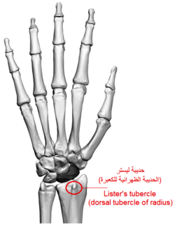 Left hand. Lister's tubercle shown. | |
| Details | |
| Identifiers | |
| Latin | tuberculum dorsale |
| TA98 | A02.4.05.017 |
| TA2 | 1226 |
| FMA | 23527 |
| Anatomical terminology | |
Lister's tubercle or dorsal tubercle of radius is a bony prominence located at the distal end of the radius. It is palpable on the dorsum of the wrist.
YouTube Encyclopedic
-
1/3Views:44 1861 5785 924
-
How to palpate the carpal bones in the wrist
-
Radius Anatomy and Osteology with Clinical Correlations
-
The Radius and Ulna Bone → Anatomy and Bony landmarks video || By: Kinesiology Kris
Transcription
Structure
Lister's tubercle is found on the dorsal distal radius.[1] It varies in size and shape significantly.[2][3] It can range from around 2 to 6 mm in height (averaging 3 mm), and around 6 to 26 mm in length (averaging 13 mm).[2][3]
Function
Lister's tubercle serves as a pulley for the tendon of extensor pollicis longus, which wraps around the medial side and takes a 45° turn.[1][2][4]
Clinical significance
Lister's tubercle is used as a useful landmark during wrist arthroscopy and other wrist surgery.[1][5] It is palpable on the dorsum of the wrist.[1] It is often difficult to clearly distinguish with radiography.[1]
Hyperextension of the wrist can lead to fracture of Lister's tubercle, as pressure is increased from the extensor pollicis longus tendon.[6] An "island-shaped" fracture can also expose the tendon to a rough edge and lead to tendon rupture (usually long after the initial fracture).[7]
References
- ^ a b c d e Chan, Wan Ying; Chong, Le Roy (2017-12-01). "Anatomical Variants of Lister's Tubercle: A New Morphological Classification Based on Magnetic Resonance Imaging". Korean Journal of Radiology. 18 (6): 957–963. doi:10.3348/kjr.2017.18.6.957. ISSN 1229-6929. PMC 5639161. PMID 29089828.
- ^ a b c Clement, Hans; Pichler, Wolfgang; Nelson, David; Hausleitner, Lisa; Tesch, Norbert Peter; Grechenig, Wolfgang (December 2008). "Morphometric Analysis of Lister's Tubercle and Its Consequences on Volar Plate Fixation of Distal Radius Fractures". The Journal of Hand Surgery. 33 (10): 1716–1719. doi:10.1016/j.jhsa.2008.08.012. ISSN 0363-5023. PMID 19084168.
- ^ a b PICHLER, W.; WINDISCH, G.; SCHAFFLER, G.; RIENMÜLLER, R.; GRECHENIG, W. (2009-10-21). "Computer tomography aided 3D analysis of the distal dorsal radius surface and the effects on volar plate osteosynthesis". Journal of Hand Surgery (European Volume). 34 (5): 598–602. doi:10.1177/1753193409101471. PMID 19959446. S2CID 45229076.
- ^ "Wheeless Online". Wheeless' Textbook of Orthopaedics. Retrieved 27 April 2014.
- ^ Ağır, İsmail; Aytekin, Mahmut Nedim; Küçükdurmaz, Fatih; Gökhan, Servan; Çavuş, Umut Yücel (2014-04-04). "Anatomical Localization of Lister's Tubercle and its Clinical and Surgical Importance". The Open Orthopaedics Journal. 8 (1): 74–77. doi:10.2174/1874325001408010074. ISSN 1874-3250. PMC 4023390. PMID 24843388.
- ^ Stahl, Shalom; Wolff, Thomas W. (May 1988). "Delayed rupture of the extensor pollicis longus tendon after nonunion of a fracture of the dorsal radial tubercle". The Journal of Hand Surgery. 13 (3): 338–341. doi:10.1016/s0363-5023(88)80004-2. ISSN 0363-5023. PMID 3379265.
- ^ Cha, Soo Min; Shin, Hyun Dae; Lee, Soong Hyun (October 2018). ""Island-shape" Fractures of Lister's tubercle have an increased risk of delayed extensor pollicis longus rupture in distal radial fractures". Injury. 49 (10): 1816–1821. doi:10.1016/j.injury.2018.08.019. ISSN 0020-1383. PMID 30154020. S2CID 52112660.