
| Lamina terminalis | |
|---|---|
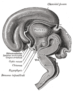 Median sagittal section of brain of human embryo of three months. (Lamina terminalis labeled at center left.) | |
 Median sagittal section of brain of human embryo of four months. (Lamina terminalis labeled at center right.) | |
| Details | |
| Identifiers | |
| Latin | lamina terminalis |
| NeuroNames | 208 |
| TA98 | A14.1.08.419 |
| TA2 | 5776 |
| FMA | 61975 |
| Anatomical terms of neuroanatomy | |
The median portion of the wall of the forebrain consists of a thin lamina, the lamina terminalis, which stretches from the interventricular foramen (foramen of Monro) to the recess at the base of the optic stalk (optic nerve) and contains the vascular organ of the lamina terminalis, which regulates the osmotic concentration of the blood. The lamina terminalis is immediately anterior to the tuber cinereum; together they form the pituitary stalk.
The lamina terminalis can be opened via endoscopic neurosurgery in an attempt to create a path that cerebrospinal fluid can flow through when a person has hydrocephalus and when it is not possible to perform an endoscopic third ventriculostomy,[1] but the effectiveness of this technique is not certain.[2]
This is the rostral end (tip) of the neural tube (embryological central nervous system) in the early weeks of development. Failure of the lamina terminalis to close properly at this stage of development will result in anencephaly or meroencephaly.
YouTube Encyclopedic
-
1/3Views:1 4076 1973 564
-
Human Brain Circumventricular Organs 3 - Organum Vasculosum LT - Sanjoy Sanyal
-
Subfrontal trans-lamina terminalis approach to a third ventricular craniopharyngioma
-
aneurysm surgery: clipping of acom aneurysm; fenestration of lamina terminalis
Transcription
Additional images
-
 Mesal aspect of a brain sectioned in the median sagittal plane.
 -
 The pituitary gland in position. Shown in sagittal section.
 -
 Cerebrum. Inferior view. Deep dissection


See also
References
![]() This article incorporates text in the public domain from page 816 of the 20th edition of Gray's Anatomy (1918)
This article incorporates text in the public domain from page 816 of the 20th edition of Gray's Anatomy (1918)
- ^ Oertel, J. M.; Vulcu, S; Schroeder, H. W.; Konerding, M. A.; Wagner, W; Gaab, M. R. (2010). "Endoscopic transventricular third ventriculostomy through the lamina terminalis". Journal of Neurosurgery. 113 (6): 1261–9. doi:10.3171/2010.6.JNS09491. PMID 20707616.
- ^ Komotar, R. J.; Hahn, D. K.; Kim, G. H.; Starke, R. M.; Garrett, M. C.; Merkow, M. B.; Otten, M. L.; Sciacca, R. R.; Connolly Jr, E. S. (2009). "Efficacy of lamina terminalis fenestration in reducing shunt-dependent hydrocephalus following aneurysmal subarachnoid hemorrhage: A systematic review. Clinical article". Journal of Neurosurgery. 111 (1): 147–54. doi:10.3171/2009.1.JNS0821. PMID 19284236.
- ^ Carlson, Neil R.; Birkett, Melissa A. (2017). Physiology of Behavior (12 ed.). Ingestive Behavior: Pearson. p. 370. ISBN 9780134320823.