
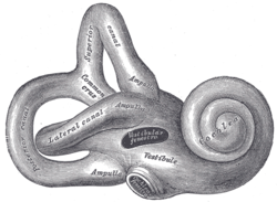

The bony labyrinth (also osseous labyrinth or otic capsule) is the rigid, bony outer wall of the inner ear in the temporal bone. It consists of three parts: the vestibule, semicircular canals, and cochlea. These are cavities hollowed out of the substance of the bone, and lined by periosteum. They contain a clear fluid, the perilymph, in which the membranous labyrinth is situated.
A fracture classification system in which temporal bone fractures detected by computed tomography are delineated based on disruption of the otic capsule has been found to be predictive for complications of temporal bone trauma such as facial nerve injury, sensorineural deafness and cerebrospinal fluid otorrhea. On radiographic images, the otic capsule is the densest portion of the temporal bone.[1][2]
In otospongiosis, a leading cause of adult-onset hearing loss, the otic capsule is exclusively affected. This area normally undergoes no remodeling in adult life and is extremely dense. With otospongiosis, the normally dense enchondral bone is replaced by Haversian bone, a spongy and vascular matrix that results in sensorineural hearing loss due to compromise of the conductive capacity of the inner ear ossicles. This results in hypodensity on CT, with the portion first affected usually being the fissula ante fenestram.[3]
The bony labyrinth is studied in paleoanthropology as it is a good indicator for distinguishing Neanderthals and modern humans.[4][5][6][7]
YouTube Encyclopedic
-
1/5Views:66 7222 4751 824496 49016 585
-
Labyrinth: Structure and inner ear function (preview) - Human Anatomy | Kenhub
-
Bony Labyrinth Of Internal Ear #shorts #Anatomy #mbbs #education
-
Anatomy of the Bony Labrynth
-
Special Senses | Inner Ear Anatomy
-
Professor Long - Ear Anatomy 2, Inner Ear
Transcription
References
- ^ Little, S. C.; Kesser, B. W. (2006). "Radiographic classification of temporal bone fractures: Clinical predictability using a new system". Archives of Otolaryngology–Head & Neck Surgery. 132 (12): 1300–4. doi:10.1001/archotol.132.12.1300. PMID 17178939.
- ^ Brodie, H. A.; Thompson, T. C. (1997). "Management of complications from 820 temporal bone fractures". The American Journal of Otology. 18 (2): 188–97. PMID 9093676.
- ^ Ho, Mai-Lan; Eisenberg, Ronald L (2014). Neuroradiology signs. New York: McGraw-Hill Medical. ISBN 978-0-07-180432-5. OCLC 1073561197.
- ^ Hublin, Jean-Jacques; Spoor, Fred; Braun, Marc; Zonneveld, Frans; Condemi, Silvana (1996). "A late Neanderthal associated with Upper Palaeolithic artefacts". Nature. 381 (6579): 224–226. Bibcode:1996Natur.381..224H. doi:10.1038/381224a0. ISSN 1476-4687. OCLC 8520954555. PMID 8622762. S2CID 4370339.
- ^ Coutinho-Nogueira, Dany; Coqueugniot, Hélène; Santos, Frédéric; Tillier, Anne-marie (20 August 2021). "The bony labyrinth of Qafzeh 25 Homo sapiens from Israel" (PDF). Archaeological and Anthropological Sciences. 13 (9): 151. doi:10.1007/s12520-021-01377-2. ISSN 1866-9565. S2CID 237219305. Archived (PDF) from the original on Dec 21, 2023.
- ^ Spoor, Fred; Hublin, Jean-Jacques; Braun, Marc; Zonneveld, Frans (16 February 2003). "The bony labyrinth of Neanderthals". Journal of Human Evolution. 44 (2): 141–165. doi:10.1016/s0047-2484(02)00166-5. ISSN 0047-2484. PMID 12662940 – via WorldCat and Elsevier Science Direct.
- ^ Stoessel, Alexander; David, Romain; Gunz, Philipp; Schmidt, Tobias; Spoor, Fred; Hublin, Jean-Jacques (2016-10-11). "Morphology and function of Neandertal and modern human ear ossicles". Proceedings of the National Academy of Sciences. 113 (41): 11489–11494. doi:10.1073/pnas.1605881113. ISSN 0027-8424. PMC 5068335. PMID 27671643.
![]() This article incorporates text in the public domain from page 1047 of the 20th edition of Gray's Anatomy (1918)
This article incorporates text in the public domain from page 1047 of the 20th edition of Gray's Anatomy (1918)
| Authority control databases: National |
|---|