
| Axillary vein | |
|---|---|
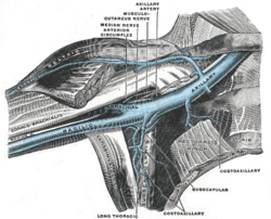 Anterior view of right upper limb and thorax - axillary vein and the distal part of the basilic vein and cephalic vein. | |
| Details | |
| Drains from | Axilla |
| Source | Basilic vein, brachial veins, cephalic vein |
| Drains to | Subclavian vein |
| Artery | Axillary artery |
| Identifiers | |
| Latin | vena axillaris |
| MeSH | D001367 |
| TA98 | A12.3.08.005 |
| TA2 | 4963 |
| FMA | 13329 |
| Anatomical terminology | |
In human anatomy, the axillary vein is a large blood vessel that conveys blood from the lateral aspect of the thorax, axilla (armpit) and upper limb toward the heart. There is one axillary vein on each side of the body.
YouTube Encyclopedic
-
1/3Views:296 879177 0222 710
-
Upper Limb Arteries - Arm and Forearm - 3D Anatomy Tutorial
-
Upper Limb Veins - 3D Anatomy Tutorial
-
Ultrasound Guidance & Steps for Axillary Vein Central Venous Access
Transcription
Structure
Its origin is at the lower margin of the teres major muscle and a continuation of the brachial vein.[1]
This large vein is formed by the brachial vein and the basilic vein.[2] At its terminal part, it is also joined by the cephalic vein.[3] Other tributaries include the subscapular vein, circumflex humeral vein, lateral thoracic vein and thoraco-acromial vein.[4] It terminates at the lateral margin of the first rib, at which it becomes the subclavian vein.[1]
It is accompanied along its course by a similarly named artery, the axillary artery, which lies laterally to the axillary vein.[5]
Additional images
-
 Intercostal nerves, the superficial muscles having been removed.
 -
 Axillary vein
 -
 Axillary vein

References
- ^ a b Baker, Champ L.; Baker, Champ L. (January 1, 2009), Wilk, Kevin E.; Reinold, Michael M.; Andrews, James R. (eds.), "CHAPTER 27 - Neurovascular Compression Syndromes of the Shoulder", The Athlete's Shoulder (Second Edition), Philadelphia: Churchill Livingstone, pp. 325–335, doi:10.1016/b978-044306701-3.50030-x, ISBN 978-0-443-06701-3, retrieved November 3, 2020
- ^ Moore, Keith L. et al. (2010) Clinically Oriented Anatomy, 6th Ed, p.718
- ^ Moore, Keith L. et al. (2010) Clinically Oriented Anatomy, 6th Ed, p.718
- ^ Moore, Keith L. et al. (2010) Clinically Oriented Anatomy, 6th Ed, fig.6.16
- ^ Gray, Andrew T., ed. (January 1, 2019), "Chapter 32 - Infraclavicular Block", Atlas of Ultrasound-Guided Regional Anesthesia (Third Edition), Elsevier, pp. 93–103, doi:10.1016/b978-0-323-50951-0.00032-3, ISBN 978-0-323-50951-0, S2CID 382483, retrieved November 3, 2020
External links
- lesson3axillaryart&vein at The Anatomy Lesson by Wesley Norman (Georgetown University)
This cardiovascular system article is a stub. You can help Wikipedia by expanding it. |