To install click the Add extension button. That's it.
The source code for the WIKI 2 extension is being checked by specialists of the Mozilla Foundation, Google, and Apple. You could also do it yourself at any point in time.
How to transfigure the Wikipedia
Would you like Wikipedia to always look as professional and up-to-date? We have created a browser extension. It will enhance any encyclopedic page you visit with the magic of the WIKI 2 technology.
Try it — you can delete it anytime.
Install in 5 seconds
Yep, but later
4,5
Kelly Slayton
Congratulations on this excellent venture… what a great idea!
Alexander Grigorievskiy
I use WIKI 2 every day and almost forgot how the original Wikipedia looks like.
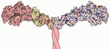
Part of the myosin II structure. Atoms in the heavy chain are colored pink (on the left-hand side); atoms in the light chains are colored faded-orange and faded-yellow (also on the left-hand side).
The first myosin (M2) to be discovered was in 1864 by Wilhelm Kühne. Kühne had extracted a viscous protein from skeletal muscle that he held responsible for keeping the tension state in muscle. He called this protein myosin.[3][4] The term has been extended to include a group of similar ATPases found in the cells of both striated muscle tissue and smooth muscle tissue.
Following the discovery in 1973 of enzymes with myosin-like function in Acanthamoeba castellanii, a global range of divergent myosin genes have been discovered throughout the realm of eukaryotes.[5]
Although myosin was originally thought to be restricted to muscle cells (hence myo-(s) + -in), there is no single "myosin"; rather it is a very large superfamily of genes whose protein products share the basic properties of actin binding, ATP hydrolysis (ATPase enzyme activity), and force transduction. Virtually all eukaryotic cells contain myosin isoforms. Some isoforms have specialized functions in certain cell types (such as muscle), while other isoforms are ubiquitous. The structure and function of myosin is globally conserved across species, to the extent that rabbit muscle myosin II will bind to actin from an amoeba.[6]
YouTube Encyclopedic
1/5
Views:
968 421
4 283
125 382
44 678
329 542
Myosin and actin | Circulatory system physiology | NCLEX-RN | Khan Academy
Myosins and its three class | Proteolytic Cleavage
Muscle Myosin
Thick Filaments and Myosin Structure
Musculoskeletal System | Sarcomere Structure: Actin & Myosin
Transcription
What I want to do in this video
is try to understand how
two proteins can interact with
each other in conjunction with
ATP to actually produce
mechanical motion.
And the reason why I want to
do this-- one, it occurs
outside of muscle cells as well,
but this is really going
to be the first video on really
how muscles work.
And then we'll talk about how
nerves actually stimulate
muscles to work.
So it'll all build up
from this video.
So what I've done here is I've
copy and pasted two images of
proteins from Wikipedia.
This is myosin.
It's actually myosin II because
you actually have two
strands of the myosin protein.
They're interwound around each
other so you can see it's this
very complex looking protein or
enzyme, however you want to
talk about it.
I'll tell you why it's called
an enzyme-- because it
actually helps react ATP into
ADP and phosphate groups.
So that's why it's
called an ATPase.
It's a subclass of the
ATPase enzymes.
This right here is actin.
What we're going to see in
this video is how myosin
essentially uses the ATP to
essentially crawl along.
You can almost view it as an
actin rope and that's what
creates mechanical energy.
So let me draw it.
I'll actually draw it on
this actin right here.
So let's say we have one
of these myosin heads.
So when I say a myosin head,
this is one of the myosin
heads right here and then it's
connected, it's interwound,
it's woven around.
This is the other one and it
winds around that way.
Now let's just say we're
just dealing with one
of the myosin heads.
Let's say it's in
this position.
Let me see how well
I can draw it.
Let's say it starts off in a
position that looks like that
and then this is kind of the
tail part that connects to
some other structural and we'll
talk about that in more
detail, but this is my myosin
head right there in its
starting position, not
doing anything.
Now, ATP can come along and bond
to this myosin head, this
enzyme, this protein,
this ATPase enzyme.
So let me draw some ATP.
So ATP comes along and bonds
to this guy right here.
Let's say that's the-- and it's
not going to be this big
relative to the protein,
but this is just to
give you the idea.
So soon as the ATP binds to its
appropriate site on this
enzyme or protein, the enzyme,
it detaches from the actin.
So let me write this down.
So one, ATP binds to myosin
head and as soon as that
happens, that causes the myosin
to release actin.
So that's step one.
So I start it off with this guy
just touching the actin,
the ATP comes, and
it gets released.
So in the next step-- so after
that step, it's going to look
something like this--
and I want to draw
it in the same place.
After the next step,
it's going to look
something like this.
It will have released.
So now it looks something like
that and you have the ATP
attached to it still.
I know it might be a little
bit convoluted when I keep
writing over the same thing,
but you have the
ATP attached to it.
Now the next step-- the ATP
hydrolizes, the phosphate gets
pulled off of it.
This is an ATPase enzyme.
That's what it does.
Let me write that down.
And what that does, that
releases the energy to cock
this myosin protein into kind
of a high energy state.
So let me do step two.
This thing-- it gets
hydrolized.
It releases energy.
We know that ATP is the energy
currency of biological
systems. So it releases
energy.
I'm drawing it as a little spark
or explosion, but you
can really imagine it's changing
the confirmation of--
it kind of spring-loads this
protein right here to go into
a state so it's ready to
crawl along the myosin.
So in step two-- plus energy,
energy and then this-- you can
say it cocks the myosin
protein or
enzyme to high energy.
You can imagine it winds the
spring, or loads the spring.
And confirmation for proteins
just mean shape.
So step two-- what happens is
the phosphate group gets--
they're still attached, but
it gets detached from
the rest of the ATP.
So that becomes ADP and that
energy changes the
confirmation so that this
protein now goes into a
position that looks like this.
So this is where we end up
at the end of step two.
Let me make sure
I do it right.
So at the end of step
two, it might look
something like this.
So the end of step two,
the protein looks
something like this.
This is in its cocked
position.
It has a lot of energy
right now.
It's wound up in
this position.
You still have your ADP.
You still have your-- that's
your adenosine and let's say
you have your two phosphate
groups on the ADP and you
still have one phosphate
group right there.
Now, when that phosphate group
releases-- so let me write
this as step three.
Remember, when we started, we
were just sitting here.
The ATP binds on step one--
actually, it does definitely
bind, at the end of step one,
that causes the myosin protein
to get released.
Then after step one, we
naturally have step two.
The ATP hydrolyzes into
ADP phosphate.
That releases energy and that
allows the myosin protein to
get cocked into this high energy
position and kind of
attach, you can think of
it, to the next rung
of our actin filament.
Now we're in a high
energy state.
In step three, the phosphate
releases.
The phosphate is released from
myosin in step three.
That's step three right there.
That's a phosphate group
being released.
And what this does is, this
releases that energy of that
cocked position and it causes
this myosin protein
to push on the actin.
This is the power stroke, if
you imagine in an engine.
This is what's causing the
mechanical movement.
So when the phosphate group is
actually released-- remember,
the original release
is when you take
ATP to ADP in a phosphate.
That put it in this
spring-loaded position.
When the phosphate releases it,
this releases the spring.
And what that does is it pushes
on the actin filament.
So you could view this
as the power stroke.
We're actually creating
mechanical energy.
So depending on which one you
want to view as fixed-- if you
view the actin as fixed,
whatever myosin is attached to
it would move to the left.
If you imagine the myosin being
fixed, the actin and
whatever it's attached to
would move to the right,
either way.
But this is where
we fundamentally
get the muscle action.
And then step four-- you
have the ADP released.
And then we're exactly where
we were before we did step
one, except we're just one rung
further to the left on
the actin molecule.
So to me, this is
pretty amazing.
We actually are seeing how ATP
energy can be used to-- we're
going from chemical energy
or bond energy in ATP to
mechanical energy.
For me, that's amazing because
when I first learned about
ATP-- people say, you use ATP to
do everything in your cells
and contract muscles.
Well, gee, how do you go from
bond energy to actually
contracting things, to actually
doing what we see in
our everyday world as
mechanical energy?
And this is really where
it all occurs.
This is really the core issue
that's going on here.
And you have to say, well, gee,
how this thing change
shape and all that?
And you have to remember,
these proteins, based on
what's bonded to it and
what's not bonded to
it, they change shape.
And some of those shapes take
more energy to attain, and
then if you do the right things,
that energy can be
released and then it can
push another protein.
But I find this just
fascinating.
And now we can build up from
this actin and myosin
interactions to understand how
muscles actually work.
Structure and functions
Domains
Most myosin molecules are composed of a head, neck, and tail domain.
The head domain binds the filamentous actin, and uses ATPhydrolysis to generate force and to "walk" along the filament towards the barbed (+) end (with the exception of myosin VI, which moves towards the pointed (-) end).
the neck domain acts as a linker and as a lever arm for transducing force generated by the catalytic motor domain. The neck domain can also serve as a binding site for myosin light chains which are distinct proteins that form part of a macromolecular complex and generally have regulatory functions.
The tail domain generally mediates interaction with cargo molecules and/or other myosin subunits. In some cases, the tail domain may play a role in regulating motor activity.
Multiple myosin II molecules generate force in skeletal muscle through a power stroke mechanism fuelled by the energy released from ATP hydrolysis.[7] The power stroke occurs at the release of phosphate from the myosin molecule after the ATP hydrolysis while myosin is tightly bound to actin. The effect of this release is a conformational change in the molecule that pulls against the actin. The release of the ADP molecule leads to the so-called rigor state of myosin.[8] The binding of a new ATP molecule will release myosin from actin. ATP hydrolysis within the myosin will cause it to bind to actin again to repeat the cycle. The combined effect of the myriad power strokes causes the muscle to contract.
The wide variety of myosin genes found throughout the eukaryotic phyla were named according to different schemes as they were discovered. The nomenclature can therefore be somewhat confusing when attempting to compare the functions of myosin proteins within and between organisms.
Skeletal muscle myosin, the most conspicuous of the myosin superfamily due to its abundance in muscle fibers, was the first to be discovered. This protein makes up part of the sarcomere and forms macromolecular filaments composed of multiple myosin subunits. Similar filament-forming myosin proteins were found in cardiac muscle, smooth muscle, and nonmuscle cells. However, beginning in the 1970s, researchers began to discover new myosin genes in simple eukaryotes[5] encoding proteins that acted as monomers and were therefore entitled Class I myosins. These new myosins were collectively termed "unconventional myosins"[9] and have been found in many tissues other than muscle. These new superfamily members have been grouped according to phylogenetic relationships derived from a comparison of the amino acid sequences of their head domains, with each class being assigned a Roman numeral[10][11][12][13] (see phylogenetic tree). The unconventional myosins also have divergent tail domains, suggesting unique functions.[14] The now diverse array of myosins likely evolved from an ancestral precursor (see picture).
Analysis of the amino acid sequences of different myosins shows great variability among the tail domains, but strong conservation of head domain sequences. Presumably this is so the myosins may interact, via their tails, with a large number of different cargoes, while the goal in each case – to move along actin filaments – remains the same and therefore requires the same machinery in the motor. For example, the human genome contains over 40 different myosin genes.
These differences in shape also determine the speed at which myosins can move along actin filaments. The hydrolysis of ATP and the subsequent release of the phosphate group causes the "power stroke", in which the "lever arm" or "neck" region of the heavy chain is dragged forward. Since the power stroke always moves the lever arm by the same angle, the length of the lever arm determines the displacement of the cargo relative to the actin filament. A longer lever arm will cause the cargo to traverse a greater distance even though the lever arm undergoes the same angular displacement – just as a person with longer legs can move farther with each individual step. The velocity of a myosin motor depends upon the rate at which it passes through a complete kinetic cycle of ATP binding to the release of ADP.
Myosin classes
Myosin I
Myosin I, a ubiquitous cellular protein, functions as monomer and functions in vesicle transport.[15] It has a step size of 10 nm and has been implicated as being responsible for the adaptation response of the stereocilia in the inner ear.[16]
Myosin II (also known as conventional myosin) is the myosin type responsible for producing muscle contraction in muscle cells in most animal cell types. It is also found in non-muscle cells in contractile bundles called stress fibers.[17]
Myosin II contains two heavy chains, each about 2000 amino acids in length, which constitute the head and tail domains. Each of these heavy chains contains the N-terminal head domain, while the C-terminal tails take on a coiled-coil morphology, holding the two heavy chains together (imagine two snakes wrapped around each other, as in a caduceus). Thus, myosin II has two heads. The intermediate neck domain is the region creating the angle between the head and tail.[18] In smooth muscle, a single gene (MYH11)[19]) codes for the heavy chains myosin II, but splice variants of this gene result in four distinct isoforms.[18]
It also contains 4 myosin light chains (MLC), resulting in 2 per head, weighing 20 (MLC20) and 17 (MLC17) kDa.[18] These bind the heavy chains in the "neck" region between the head and tail.
Self-inhibition of Myosin II.[20][21][22] The movie begins with Myosin II in the 10S conformation with a folded tail domain, the blocked head and free head.[23][24] The movie schematically depicts tail unfolding and the resulting active 6S confirmation followed by tail folding back to the 10S conformation.[25][26] The illustration is conceptual: transitory states and diffusive motions associated with folding/unfolding are not shown.[27]The MLC20 is also known as the regulatory light chain and actively participates in muscle contraction.[18]
The MLC17 is also known as the essential light chain.[18] Its exact function is unclear, but is believed to contribute to the structural stability of the myosin head along with MLC20.[18] Two variants of MLC17 (MLC17a/b) exist as a result of alternative splicing at the MLC17 gene.[18]
In muscle cells, the long coiled-coil tails of the individual myosin molecules can auto-inhibit active function in the 10S conformation or upon phosphorylation, change to the 6S conformation and join, forming the thick filaments of the sarcomere.[28][29] The force-producing head domains stick out from the side of the thick filament, ready to walk along the adjacent actin-based thin filaments in response to the proper chemical signals and may be in either auto-inhibited or active conformation. The balance/transition between active and inactive states is subject to extensive chemical regulation.
Crystal structure of myosin V motor with essential light chain – nucleotide-free
Myosin IV
Myosin IV has a single IQ motif and a tail that lacks any coiled-coil forming sequence. It has homology similar to the tail domains of Myosin VII and XV.[32]
Myosin V
Myosin V is an unconventional myosin motor, which is processive as a dimer and has a step size of 36 nm.[33] It translocates (walks) along actin filaments traveling towards the barbed end (+ end) of the filaments. Myosin V is involved in the transport of cargo (e.g. RNA, vesicles, organelles, mitochondria) from the center of the cell to the periphery, but has been furthermore shown to act like a dynamic tether, retaining vesicles and organelles in the actin-rich periphery of cells.[34][35] A recent single molecule in vitro reconstitution study on assembling actin filaments suggests that Myosin V travels farther on newly assembling (ADP-Pi rich) F-actin, while processive runlengths are shorter on older (ADP-rich) F-actin.[36]
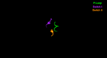
A ribbon diagram of the Myosin V molecular motor[37] pseudo-colored to illustrate major subdomains. In the interest of visual clarity, important loops (which are often labeled separately in the literature) are not singled out. This perspective highlights the nucleotide-binding site and the separation of the U50 and L50 subdomains which form the actin-binding site cleft.
The Myosin V motor head can be subdivided into the following functional regions:[37]
Nucleotide-binding site - These elements together coordinate di-valent metal cations (usually magnesium) and catalyze hydrolysis:
Switch I - This contains a highly conserved SSR motif. Isomerizes in the presence of ATP.
Switch II - This is the Kinase-GTPase version of the Walker B motif DxxG. Isomerizes in the presence of ATP.
P-loop - This contains the Walker A motif GxxxxGK(S,T). This is the primary ATP binding site.
Transducer - The seven β-strands that underpin the motor head's structure.[38]
U50 and L50 - The Upper (U50) and Lower (L50) domains are each around 50kDa. Their spatial separation[39] forms a cleft critical for binding to actin and some regulatory compounds.
SH1 helix and Relay - These elements together provide an essential mechanism for coupling the enzymatic state of the motor domain to the powerstroke-producing region (converter domain, lever arm, and light chains).[40][41]
Converter - This converts a change of conformation in the motor head to an angular displacement of the lever arm (in most cases reinforced with light chains).[41]
Myosin VI
State of myosin VI from PDB 2V26 before the power stroke [42]
Myosin VI is an unconventional myosin motor, which is primarily processive as a dimer, but also acts as a nonprocessive monomer. It walks along actin filaments, travelling towards the pointed end (- end) of the filaments.[43] Myosin VI is thought to transport endocytic vesicles into the cell.[44]
Myosin VIII is a plant-specific myosin linked to cell division;[47] specifically, it is involved in regulating the flow of cytoplasm between cells[48] and in the localization of vesicles to the phragmoplast.[49]
Myosin IX
Myosin IX is a group of single-headed motor proteins. It was first shown to be minus-end directed,[50] but a later study showed that it is plus-end directed.[51] The movement mechanism for this myosin is poorly understood.
Myosin X
Myosin X is an unconventional myosin motor, which is functional as a dimer. The dimerization of myosin X is thought to be antiparallel.[52] This behavior has not been observed in other myosins. In mammalian cells, the motor is found to localize to filopodia. Myosin X walks towards the barbed ends of filaments. Some research suggests it preferentially walks on bundles of actin, rather than single filaments.[53] It is the first myosin motor found to exhibit this behavior.
Myosin XI
Myosin XI directs the movement of organelles such as plastids and mitochondria in plant cells.[54] It is responsible for the light-directed movement of chloroplasts according to light intensity and the formation of stromules interconnecting different plastids. Myosin XI also plays a key role in polar root tip growth and is necessary for proper root hair elongation.[55] A specific Myosin XI found in Nicotiana tabacum was discovered to be the fastest known processive molecular motor, moving at 7μm/s in 35 nm steps along the actin filament.[56]
Myosin XII
Myosin XIII
Myosin XIV
This myosin group has been found in the Apicomplexa phylum.[57] The myosins localize to plasma membranes of the intracellular parasites and may then be involved in the cell invasion process.[58]
This myosin is also found in the ciliated protozoan Tetrahymena thermaphila. Known functions include: transporting phagosomes to the nucleus and perturbing the developmentally regulated elimination of the macronucleus during conjugation.
Myosin XV
Myosin XV is necessary for the development of the actin core structure of the non-motile stereocilia located in the inner ear. It is thought to be functional as a monomer.
Myosin XVI
Myosin XVII
Myosin XVIII
MYO18A A gene on chromosome 17q11.2 that encodes actin-based motor molecules with ATPase activity, which may be involved in maintaining stromal cell scaffolding required for maintaining intercellular contact.
Myosin XIX
Unconventional myosin XIX (Myo19) is a mitochondrial associated myosin motor.[59]
Myosin light chains are distinct and have their own properties. They are not considered "myosins" but are components of the macromolecular complexes that make up the functional myosin enzymes.
Paramyosin is a large, 93-115kDa muscleprotein that has been described in a number of diverse invertebrate phyla.[60] Invertebrate thick filaments are thought to be composed of an inner paramyosin core surrounded by myosin. The myosin interacts with actin, resulting in fibre contraction.[61] Paramyosin is found in many different invertebrate species, for example, Brachiopoda, Sipunculidea, Nematoda, Annelida, Mollusca, Arachnida, and Insecta.[60] Paramyosin is responsible for the "catch" mechanism that enables sustained contraction of muscles with very little energy expenditure, such that a clam can remain closed for extended periods.
^Goodson HV (1994). "Molecular evolution of the myosin superfamily: application of phylogenetic techniques to cell biological questions". Society of General Physiologists Series. 49: 141–157. PMID7939893.
^Matsuoka R, Yoshida MC, Furutani Y, Imamura S, Kanda N, Yanagisawa M, et al. (April 1993). "Human smooth muscle myosin heavy chain gene mapped to chromosomal region 16q12". American Journal of Medical Genetics. 46 (1): 61–67. doi:10.1002/ajmg.1320460110. PMID7684189.
^Pettersen EF, Goddard TD, Huang CC, Couch GS, Greenblatt DM, Meng EC, et al. (October 2004). "UCSF Chimera--a visualization system for exploratory research and analysis". Journal of Computational Chemistry. 25 (13): 1605–1612. doi:10.1002/jcc.20084. PMID15264254.
^Hammer JA, Sellers JR (December 2011). "Walking to work: roles for class V myosins as cargo transporters". Nature Reviews. Molecular Cell Biology. 13 (1): 13–26. doi:10.1038/nrm3248. PMID22146746. S2CID11853457.
^Kull FJ, Vale RD, Fletterick RJ (November 1998). "The case for a common ancestor: kinesin and myosin motor proteins and G proteins". Journal of Muscle Research and Cell Motility. 19 (8): 877–886. doi:10.1023/a:1005489907021. PMID10047987. S2CID25508217.
^Inoue A, Saito J, Ikebe R, Ikebe M (April 2002). "Myosin IXb is a single-headed minus-end-directed processive motor". Nature Cell Biology. 4 (4): 302–306. doi:10.1038/ncb774. PMID11901422. S2CID12158370.
^O'Connell CB, Mooseker MS (February 2003). "Native Myosin-IXb is a plus-, not a minus-end-directed motor". Nature Cell Biology. 5 (2): 171–172. doi:10.1038/ncb924. PMID12563277. S2CID687308.
Soldati T, Geissler H, Schwarz EC (1999). "How many is enough? Exploring the myosin repertoire in the model eukaryote Dictyostelium discoideum". Cell Biochemistry and Biophysics. 30 (3): 389–411. doi:10.1007/BF02738121. PMID10403058. S2CID13319819.
Molecular Biology of the Cell. Alberts, Johnson, Lewis, Raff, Roberts, and Walter. 4th Edition. 949–952.
http://cellimages.ascb.org/cdm4/item_viewer.php?CISOROOT=/p4041coll12&CISOPTR=101&CISOBOX=1&REC=2[dead link] Animation of a moving myosin motor protein


 Phase 1
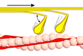 Phase 2
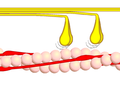 Phase 3
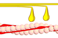 Phase 4


