
| Myoepithelial cell | |
|---|---|
 | |
| Details | |
| Identifiers | |
| Latin | myoepitheliocytus |
| TH | H2.00.02.0.03059 |
| FMA | 67799 67805, 67799 |
| Anatomical terminology | |
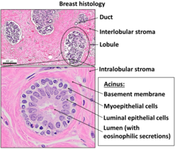
Myoepithelial cells (sometimes referred to as myoepithelium) are cells usually found in glandular epithelium as a thin layer above the basement membrane but generally beneath the luminal cells. These may be positive for alpha smooth muscle actin and can contract and expel the secretions of exocrine glands. They are found in the sweat glands, mammary glands, lacrimal glands, and salivary glands. Myoepithelial cells in these cases constitute the basal cell layer of an epithelium that harbors the epithelial progenitor. In the case of wound healing, myoepithelial cells reactively proliferate. Presence of myoepithelial cells in a hyperplastic tissue proves the benignity of the gland and, when absent, indicates cancer. Only rare cancers like adenoid cystic carcinomas contains myoepithelial cells as one of the malignant components.
It can be found in endoderm or ectoderm.[1]
YouTube Encyclopedic
-
1/1Views:159 969
-
Breast Anatomy
Transcription
Let's draw a diagram of the human breast and talk about some of the amazing functions of this organ. Now, the human breast in both males and females contains mammary glands. And in females, around the time of puberty, these mammary glands develop. In males, they usually remain undeveloped. And the main function of these glands-- and I'll just write in the name over here. These are called the mammary glands, and the main function of the mammary glands is to secrete milk to nourish the human infant. Now, these mammary glands, each of them drains toward the nipple by a little duct called a lactiferous duct. And I remember when I was expecting my first baby, and I remember thinking, where is the milk going to come out from? I don't see a hole there. And the answer is, there isn't a single hole. There are many tiny holes that are so small that you can't actually see them with the naked eye. And so these lactiferous ducts-- and I'll just label them for you here. These are the lactiferous ducts. And the lactiferous ducts empty, or drain, towards the nipple and towards that darker area of skin that we see when we look at the human breast. And that area skin is called the areola. And the reason the areola exists, is because when the newborn first comes out, newborns actually don't see very well. And in order to help them find their source of food, we have this darkened area of the breast that actually gets quite a bit darker during pregnancy. OK. So now, these mammary glands are lined with what we call myoepithelial cells. And let me just break down that word for you. Myo is a prefix that we use when we're talking about anything that has to do with muscles or anything contracts. So these are, we know, contractile cells. And they're also epithelial cells, and epithelial cells in the body are the cells that line things. So these cells, these red ones, these myoepithelial cells, are cells that both line the mammary glands and have the ability to contract to eject the milk out through the lactiferous ducts. Now, all of these structures are supported by quite a bit of connective tissue. And we're usually talking about-- in the breast the main ones are collagen and elastin. And all of these connective tissue framework comes together in these ligaments, strong ligaments that anchor the breast to the chest wall. And these ligaments are called Cooper's ligaments. In medical school, we used to remember this by saying the phrase Cooper's droopers. But after having had three children and breast fed for a long time, I don't think that's so funny anymore. Anyway, the only other thing in the breast that makes it kind of a soft and squishy organ is the adipose tissue that also forms part of the framework, or the structure, and supports actually all of these glands along with the connective tissue, the collagen and elastic. So this is our adipose tissue. And we'll see in our next video what happens, how the body knows when to cause these myoepithelial cells to contract, to eject the milk out of the mammary glands.
Markers
Myoepithelial cells are true epithelial cells positive for keratins, not to be confused with myofibroblasts which are true mesenchymal cells positive for vimentin. These cells are generally positive for alpha smooth muscle actin (αSMA), cytokeratin 5/6 and other high molecular weight cytokeratins, p63 and caldesmon. Myoepithelial cells are stellate in shape and are also known as basket cells. They lie between the basement membrane and glandular epithelium. Each cell consists of a cell body from which 4-8 processes radiate and embrace the secretory unit. Myoepithelial cells have contractile functions. They help in expelling secretions from the lumen of secretory units and facilitate the movement of saliva in salivary ducts.
Other Cancers Involving This Type of Cell

- Myoepithelioma of the head and neck - A (usually) benign tumor of the head/neck consisting of solely myoepithelial cells.
- Epithelial-myoepithelial carcinoma (of the salivary glands) - A low-grade malignant tumor composed of both neoplastic epithelial and neoplastic myoepithelial cells (a biphasic tumor).
- Epithelial-myoepithelial carcinoma of the lung- A malignant tumor composed of both epithelial and myoepithelial tissues whose pathology resemble salivary cells.
- Adenomyoepithelioma of the breast- A (usually) benign tumor of the breast composed of Myoepithelial and Adeno (glandular) cells.
- Myoepithelioma of the Breast- A usually benign or exceedingly rare malignant tumor of the breast which mimics IDC but with cells resembling adenoid cysts. If malignant, this is also known as a Myoepithelial Carcinoma.
See also
List of distinct cell types in the adult human body
References
- ^ Jules J. Berman (2009). Neoplasms: principles of development and diversity. Jones & Bartlett Learning. pp. 207–. ISBN 978-0-7637-5570-6. Retrieved 16 April 2010.
External links
- Anatomy Atlases – Microscopic Anatomy, plate 07.141 - "Axillary Sweat Gland: Myoepithelium"
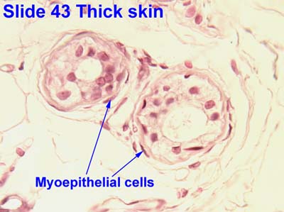
- Histology image: 43_13 at the University of Oklahoma Health Sciences Center - "thick skin"
- Histology at KUMC glands-glands09 "Simple Tubular Coiled"
- Costoff, A., Essentials of Human Physiology, archived from the original on 2015-11-20
{{citation}}: CS1 maint: bot: original URL status unknown (link)
This anatomy article is a stub. You can help Wikipedia by expanding it. |