
| Corpus cavernosum of clitoris | |
|---|---|
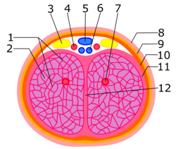 Internal anatomy of clitoral body: 1. Trabeculae of corpora cavernosa of clitoris; 2. Cavernous spaces; 3. Dorsal nerve of clitoris; 4. Dorsal artery of clitoris; 5. Superficial dorsal veins of clitoris; 6. Deep dorsal vein of clitoris; 7. Deep artery of clitoris; 8. Skin; 9. Membranous layer of subcutaneous tissue; 10. Clitoral fascia; 11. Tunica albuginea; 12. Septum of clitoris. | |
 The internal anatomy of the human vulva, with the clitoral hood and labia minora indicated as lines. | |
| Details | |
| Part of | Clitoris |
| System | Reproductive system |
| Function | Clitoral erection |
| Identifiers | |
| Latin | corpus cavernosum clitoridis |
| TA98 | A09.2.02.005 |
| TA2 | 3569 |
| FMA | 20172 |
| Anatomical terminology | |
The corpus cavernosum of clitoris is one of a pair of sponge-like regions of erectile tissue of the clitoris. It is made of a sponge-like tissue that fills with blood during erection. This is homologous to the corpus cavernosum penis. The term corpora cavernosa literally means "cave-like bodies".
YouTube Encyclopedic
-
1/3Views:286 5761 2521 628
-
02.Female Repro System. Perineum
-
Erectile Tissues M1 Perineum SDV
-
The Five Parts of the Clitoris
Transcription
Structure

The two corpora cavernosa are expandable erectile tissues of the clitoris.[1] They are joined together along their medial surfaces by an incomplete fibrous septum.[2] Each corpus cavernosum is connected to the rami of the pubis and ischium by a clitoral crus.[1][3] There is connection to the ischiocavernosus muscle.[2] Each can be up to 7 cm long in an adult.[1]
Development
The corpus cavernosum is homologous to the corpus cavernosum penis in the male. It develops from the genital tubercle in the embryo.[1]
The clitoris also has two vestibular bulbs beneath the skin of the labia minora (at the entrance to the vagina), which expand at the same time as the glans clitoridis to cap the ends of the corpora cavernosa.
Microanatomy
The corpus cavernosum is made of a sponge-like tissue. This contains irregular blood-filled spaces, lined by endothelium, and separated by connective tissue septa.
Function
The corpora cavernosa fill with blood during clitoral erection.[1] Their size increases 2 fold to 3 fold.[1] In some circumstances, release of nitric oxide precedes relaxation of the clitoral cavernosal artery and nearby muscle, in a process similar to male arousal. More blood flows in through the clitoral cavernosal artery, the pressure in the corpora cavernosa clitoridis rises. The clitoris is engorged with blood. This leads to extrusion of the glans clitoridis and enhanced sensitivity to physical contact.
Clinical significance
The corpora cavernosa can reduce in size before the menopause.[1] This can impair sexual function in some women.[1]
References
- ^ a b c d e f g h Graziottin, Alessandra; Gambini, Dania (2015). "4 - Anatomy and physiology of genital organs – women". Handbook of Clinical Neurology. Vol. 130. Elsevier. pp. 39–60. doi:10.1016/B978-0-444-63247-0.00004-3. ISBN 978-0-444-63247-0. ISSN 0072-9752. PMID 26003238.
- ^ a b Lowe, James S.; Anderson, Peter G. (2015). "17 - Female Reproductive System". Stevens & Lowe's Human Histology (4th ed.). Mosby. pp. 337–362. doi:10.1016/B978-0-7234-3502-0.00017-6. ISBN 978-0-7234-3502-0.
- ^ Gray, Henry (1918). Atlas of the Human Body. Lea & Febiger.